-
 Univers
-
 Ebooks
-
 Livres audio
-
 Presse
-
 Podcasts
-
 BD
BD
-
 Documents
-
- Cours
- Révisions
- Ressources pédagogiques
- Sciences de l’éducation
- Manuels scolaires
- Langues
- Travaux de classe
- Annales de BEP
- Etudes supérieures
- Maternelle et primaire
- Fiches de lecture
- Orientation scolaire
- Méthodologie
- Corrigés de devoir
- Annales d’examens et concours
- Annales du bac
- Annales du brevet
- Rapports de stage
La lecture à portée de main

Vous pourrez modifier la taille du texte de cet ouvrage
Découvre YouScribe en t'inscrivant gratuitement
Je m'inscrisSelf-assessment in Axial Musculoskeletal Trauma X-rays , livre ebook
Découvre YouScribe en t'inscrivant gratuitement
Je m'inscrisEn savoir plus
Vous pourrez modifier la taille du texte de cet ouvrage
En savoir plus

Description
Sujets
Informations
| Publié par | M&K Publishing |
| Date de parution | 14 août 2009 |
| Nombre de lectures | 0 |
| EAN13 | 9781907830471 |
| Langue | English |
Informations légales : prix de location à la page 0,0960€. Cette information est donnée uniquement à titre indicatif conformément à la législation en vigueur.
Extrait
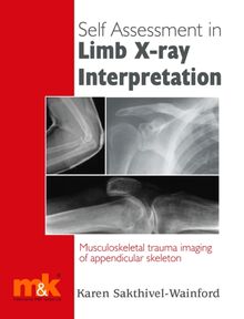
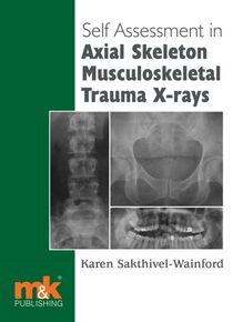
Self Assessment in Axial Skeleton Musculoskeletal Trauma X-rays
Also in this series:
Self-assessment in Limb X-ray Interpretation ISBN: 978-1-905539-13-0 2006
Self-assessment in Paediatric Musculoskeletal Trauma X-rays ISBN: 978-1-905539-34-5 2008
Forthcoming:
Self-assessment in Musculoskeletal Pathology
Other health and social care books from M K include
Routine Blood Results Explained 2/e ISBN: 978-1-905539-38-3 2008
A Guide to Research for Podiatrists ISBN: 978-1-905539-41-3 2007
The ECG Workbook ISBN: 978-1-905539-14-7 2008
Arterial Blood Gas Analysis: An easy learning guide ISBN: 978-1-905539-04-8 2008
Haemodynamic Monitoring Manipulation: An easy learning guide ISBN: 978-1-905539-46-8 2009
The Management of COPD in Primary and Secondary Care ISBN: 978-1-905539-28-4 2007
The Clinician s Guide to Chronic Disease Management for Long Term Conditions: A cognitive-behavioural approach ISBN: 978-1-905539-15-4 2008
Self Assessment in
Axial Skeleton Musculoskeletal Trauma X-rays
Karen Sakthivel-Wainford
H.D.C.R (R), PGCert, MSc Advanced Radiographer Practitioner, Leeds General Infirmary, UK
Self-assessment in Axial Skeleton Musculoskeletal Trauma X-rays Karen Sakthivel-Wainford
ISBN: 978-1-905539-47-5
First published 2009
All rights reserved. No part of this publication may be reproduced, stored in a retrieval system, or transmitted in any form or by any means, electronic, mechanical, photocopying, recording or otherwise, without either the prior permission of the publishers or a licence permitting restricted copying in the United Kingdom issued by the Copyright Licensing Agency, 90 Tottenham Court Road, London, W1T 4LP. Permissions may be sought directly from M K Publishing, phone: 01768 773030, fax: 01768 781099 or email: publishing@mkupdate.co.uk
Any person who does any unauthorised act in relation to this publication may be liable to criminal prosecution and civil claims for damages.
British Library Cataloguing in Publication Data A catalogue record for this book is available from the British Library
Notice Clinical practice and medical knowledge constantly evolve. Standard safety precautions must be followed, but, as knowledge is broadened by research, changes in practice, treatment and drug therapy may become necessary or appropriate. Readers must check the most current product information provided by the manufacturer of each drug to be administered and verify the dosages and correct administration, as well as contraindications. It is the responsibility of the practitioner, utilising the experience and knowledge of the patient, to determine dosages and the best treatment for each individual patient. Any brands mentioned in this book are as examples only and are not endorsed by the Publisher. Neither the publisher nor the authors assume any liability for any injury and/or damage to persons or property arising from this publication.
The Publisher
To contact M K Publishing write to:
M K Update Ltd The Old Bakery St. John's Street
Keswick Cumbria CA12 5AS Tel: 01768 773030 Fax: 01768 781099
publishing@mkupdate.co.uk www.mkupdate.co.uk
Designed and typeset in Usherwood Book by Mary Blood Printed in England by Reeds Printers, Penrith .
Contents
Acknowledgements
Introduction
1. Mechanisms of injury
2. Pelvic fractures
3. Reviewing cervical spine radiographs
4. Pelvic trauma
5. Hip and femur trauma
6. Cervical spine trauma
7. Dorsal and lumbar spine trauma
8. Skull, mandibular and facial trauma
9. A selection of cases
Reading list and bibliography
Index
Acknowledgements
To the Radiology departments of Leeds General Infirmary, and Wharfedale General Hospital, for their continued support and the use of radiographs in this book.
To Katy Johnson for participating in this book.
To Fiona Carmichael, for help and support when I have to lecture on mandible and facial trauma.
In memory of my beloved mother, Marjorie Wainford, who has not long left this world. Her courage and strength of mind through difficulties will always be an inspiration to me.
Introduction
Many radiographer practitioners are now continuing to expand their reporting skills from the appendicular skeleton to include the axial skeleton in trauma. Other allied professions may also be reviewing axial skeleton trauma radiographs, for instance nurse practitioners (particularly in cases of hip trauma, in our trust), physiotherapists, etc., and hence wish to increase their knowledge. This book, like the others in the series is written specifically for radiographers, be they red dotting radiographers, commenting radiographers, radiographer practitioners or nurse practitioners.
Many radiographers initially fear reviewing axial skeleton radiographs, understandably, as missing an injury may have dire consequences. But with training, audit and care this fear can be overcome; and one can look forward to the challenge of axial radiograph reporting.
The book is intended to be like sitting in on a reporting session with a radiologist or radiographer practitioner, where you are asked to report/comment on the radiograph, but also asked some questions and given feedback. It is intended to support whatever course you have done, whether it be reporting, red dotting or commenting, or to encourage you to go on that course . It can be a revision book, or help in preparation for an assessment. However you use it, I hope it will encourage you to read more and research more into musculoskeletal trauma.
As axial trauma radiographs can be difficult to review, the book starts with several chapters, to introduce or revise specific axial trauma. The first chapter discusses mechanisms of injury of major trauma and is written by Katy Johnson, a Radiographer Practitioner working at Leeds General Infirmary. This is followed by a chapter on pelvic trauma. The next chapter looks at reviewing cervical spine trauma radiographs.
Then a series of trauma cases of the axial skeleton is presented, on which you are asked to write reports, and sometimes answer a few questions (the answers are over the page). This section is divided into six chapters: trauma cases of the pelvis; of the hip and femur; the cervical spine; dorsal and lumbar spine; the skull, facial bones and mandible (15 cases in each chapter); the last chapter being 25 mixed cases. It is preferable to work your way through the book from start to finish. However, if you feel you need revision on, say, cervical spine radiographs, then you can flick to the chapter on reviewing the cervical spine and then to the cases on the cervical spine.
Each case has appropriate clinical history, although this may not be the original history in order to anonymise the case. Some of the cases may not have side markers; these may have been removed whilst removing patients details.
Note : Specific references appear at the end of Chapter 1 . There is also a general reading list/bibliography on page 267.
1.
Mechanisms of injury
Katy Johnson
In order to understand how injuries occur, we must recognise the importance of the mechanism of injury in determining injury patterns. Each situation will be different, but by obtaining an accurate assessment of how the accident happened and the mechanisms involved, certain injury patterns can be predicted. This chapter looks at some basic laws of physics and explains how these relate to the trauma setting.
There are several mechanisms which are highly predictive of serious injury due to the forces involved (Greaves, Porter and Ryan, 2001):
Fall of more than 6 metres
Pedestrian or cyclist hit by car
Death of other occupant in same vehicle
Ejection from vehicle/bike
Major vehicular deformity or significant intrusion into passenger space
Extrication time more than 20 minutes
Vehicular roll-over
Penetrating injury to head or torso
All shotgun wounds.
All of the above mechanisms share the same common factor - they arise as a direct result of significant energy transfer. But before we can appreciate how the injuries occur, we need to understand some basic laws of motion.
Basic laws of motion
Many injuries occur as a result of the transfer of Kinetic Energy (KE) - this is the energy of motion and can be defined as:
KE = 1 / 2 mv 2
where m = mass and v = velocity.
Newton s First Law states that an object which is at rest will stay at rest and an object which is in motion will stay in motion in a straight line unless acted upon by an external force. This is often referred to as the law of inertia.
Newton s Second Law builds on the first and states that force (F) is equal to the product of mass (m) and acceleration (a):
F = ma
which means that force is associated with a change in momentum, i.e. acceleration or deceleration.
Newton s Third Law states that for every action, there is an equal and opposite reaction.
This in turn brings in another basic law of motion, that energy cannot be created or destroyed, but it can change in form or be absorbed. It is this energy change and absorption that affects the human body and produces a specific pattern of injuries.
Once we understand these laws, we can relate them to the effects they have upon the human body in real life trauma situations.
Impact characteristics
Mechanical injuries are produced when two bodies collide and the kinetic energy is dissipated. There are several characteristics of the collision which influence the degree of injury to the human body (Nowak and Handford, 1999).
The total amount of energy which is transferred from one object to another plays a massive role in the damage sustained. The heavier an object is and the faster it is moving, the more kinetic energy it possesses. If you double the mass of an object, you double the kinetic energy, whereas if you double the velocity of it, four times the kinetic energy is created. Therefore, if a person who has a mass of 70kg drives his car with a velocity of 30km/hour, he will have a kineti

-
Univers
-
Ebooks
-
Livres audio
-
Presse
-
Podcasts
-
BD
-
Documents
-
Jeunesse
-
Littérature
-
Ressources professionnelles
-
Santé et bien-être
-
Savoirs
-
Education
-
Loisirs et hobbies
-
Art, musique et cinéma
-
Actualité et débat de société
-
Jeunesse
-
Littérature
-
Ressources professionnelles
-
Santé et bien-être
-
Savoirs
-
Education
-
Loisirs et hobbies
-
Art, musique et cinéma
-
Actualité et débat de société
-
Actualités
-
Lifestyle
-
Presse jeunesse
-
Presse professionnelle
-
Pratique
-
Presse sportive
-
Presse internationale
-
Culture & Médias
-
Action et Aventures
-
Science-fiction et Fantasy
-
Société
-
Jeunesse
-
Littérature
-
Ressources professionnelles
-
Santé et bien-être
-
Savoirs
-
Education
-
Loisirs et hobbies
-
Art, musique et cinéma
-
Actualité et débat de société
- Cours
- Révisions
- Ressources pédagogiques
- Sciences de l’éducation
- Manuels scolaires
- Langues
- Travaux de classe
- Annales de BEP
- Etudes supérieures
- Maternelle et primaire
- Fiches de lecture
- Orientation scolaire
- Méthodologie
- Corrigés de devoir
- Annales d’examens et concours
- Annales du bac
- Annales du brevet
- Rapports de stage

